Abdomen Protocol
-
Scanning Duration: 30 minutes for full abdomen.
Aorta Sag Prox/Mid/Distal
- Before even taking your first image, determine the correct frequency setting based on your patient.
- Proximal aorta must include diaphragmatic border, so angle the probe cranially.
- Use TGC to darken the normal aorta lumen or remove artifact. Color image: One at mid aorta, with good color adjustment (use the aorta color preset).


Aorta Sag
IVC Sag
- Obtain proximal IVC with vessel going into the heart.
- Use TGC to darken the normal IVC lumen or remove artifact.
- If the patient is too gassy at midline, you can also find your window through the ribs using a right lateral approach. Color image: One at proximal IVC, with good color filling.


IVC Sag
Left Lobe Liver Sag (Med-Lat)
- Obtain 3 stills showing the left liver with IVC/caudate lobe, aorta, and liver tip.
- Diaphragm should be in view, demarcating superior border of the left lobe. Cine: Medial to lateral, or lateral to medial.


Lt Liver Sag
Left Lobe Liver Trans (Sup-Inf)
- Obtain 3 stills showing the left liver superiorly with diaphragm, IVC/caudate lobe, and LPV.
- The round ligament separates right and left lobe, so use this as a landmark. Color image: One at LPV (red color filling).
Cine: Superior to inferior.


Lt Liver Trans
Right Lobe Liver Sag
- Take an image showing liver and right kidney interface.
- Depending on your window, angle cranially or slide superiorly until you see right liver dome. Shown in the diagram: subcostal approach angling cranially. Measurement image: From liver dome (most superior portion visible) to tip.


Rt Liver Sag
Right Lobe Liver Sag (Lat-Med)
- Obtain at least 3 stills showing liver dome, hepatic veins, and liver with right kidney or gallbladder.
- In a subcostal approach (as illustrated), your transducer is flush with the rib border. Angle cranially or underneath the ribs, slide infero-medially.
- If supine position is suboptimal, turn the patient on his left side and reevaluate. Cine: Take one cine.


Rt Liver Sag
Right Lobe Liver Trans (Sup-Inf)
- Obtain at least 3 stills showing liver dome, hepatic veins, portal vein, and liver with right kidney or gallbladder.
- In a subcostal approach (as illustrated), your transducer is flush with the rib border. Note that the notch is now facing you.
- Survey the whole right liver by starting at position 1, then moving more laterally to position 2. Cine: Take at least one cine from position 1 and/or 2.


Rt Liver Trans
Hepatic V
- Find your window subcostally in transverse, or as a second option, intercostally by right lateral approach as shown. Color image: Show the veins with good color filling (color blue).
Doppler image: Sample any one hepatic vein with adjustment to angle, baseline, speed, scale, and gain. You should optimize these parameters every time.


Hepatic Vein
MPV
- Use a right lateral approach.
- Normal velocity 20-40 cm/sec. Color image: Show the extrahepatic portal vein with good color filling (color red).
Doppler image: Sample the center of the vein with adjustment to angle, baseline, speed, scale, and gain.


Main Portal Vein
RPV
- Use a right lateral approach, scanning between the ribs. Color image: Show the intrahepatic portal vein with good color filling (color red) with either an anterior branch (color red) and/or posterior branch (color blue).
CBD
- Use a right lateral approach as a default for most patients, at the same area of MPV.
- If the patient is skinny, you can also get CBD at midline near the pancreas head.
- Zoom in the screen, and decrease gain or adjust TGC.
- Normal diameter <6 mm under the age 60, and ≤10 mm after cholecystectomy. Color image: Take one color image before your grayscale with measurement.
Measurement image: Calipers placed from inner wall to inner wall.

Common Bile Duct
Pancreas Trans (Head/Body/Tail)
- Label and take Head/Body/Tail if you have good visualization of the entire pancreas.
- Make a conscious note at this point if there is any ductal dilatation (read: "double duct sign"). Color image: Take one color image (color blue at pancreas head, red at tail).
Cine: Superior to inferior.

Pancreas Trans
Pancreas Sag (Right-Left)
- One image at pancreas head with the confluence.
- Adjust gain or TGC to darken the normal lumen of the portal confluence. Color image: Take one color image at the pancreas head with portal confluence (color blue).
Cine: Right to left.

Pancreas Sag
Gallbladder Sag Supine
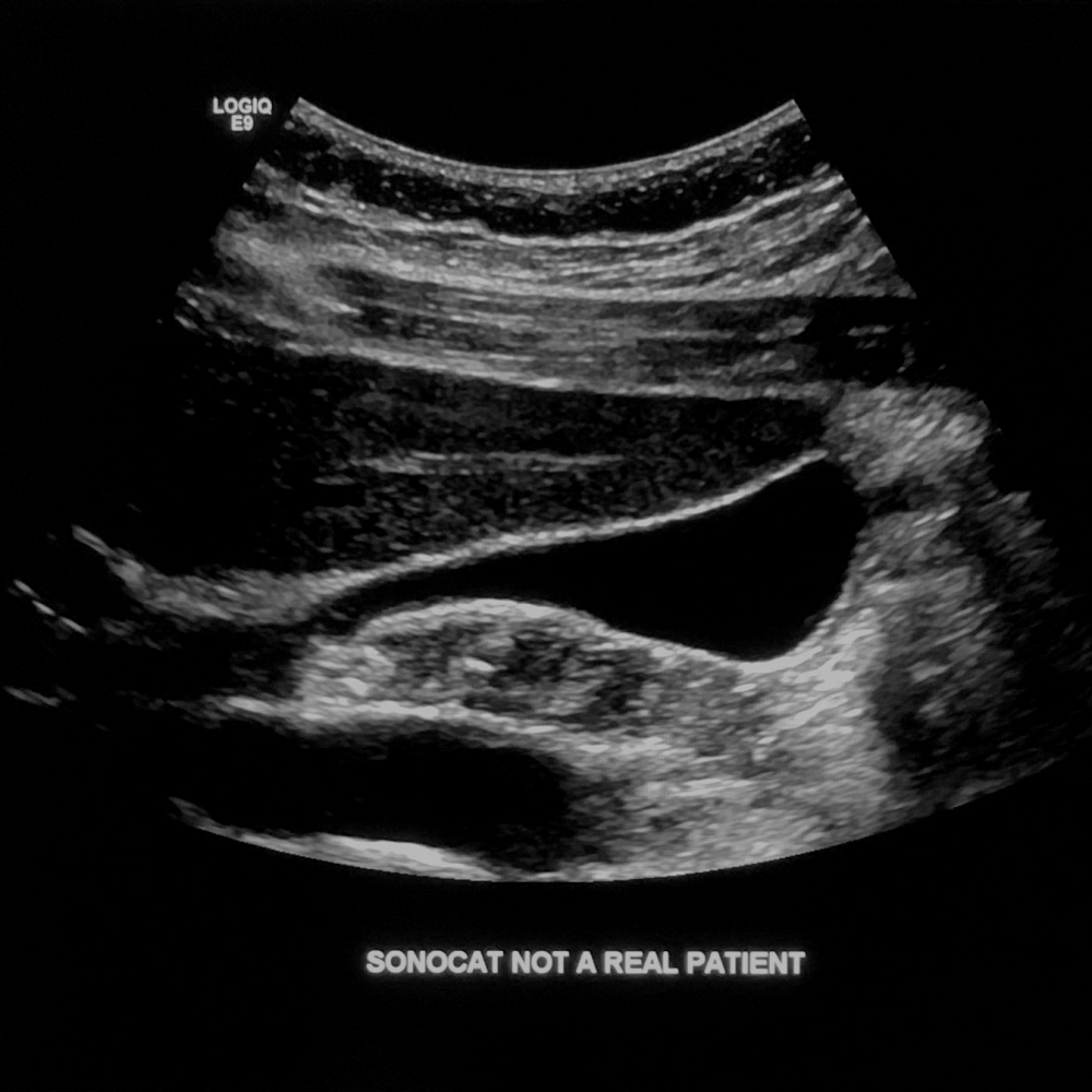
- One image showing neck, body, and fundus.
- Adjust gain or TGC, and find different windows to avoid reverberation artifact.
- No need to measure the wall if it is normal.
- Assess for sonographic Murphy's sign.
- If the gallbladder is absent, label "Gallbladder Fossa Sag". Color image: Only if wall appears thickened or >3 mm for possible inflammation, or when there are findings (e.g. mass-like lesions).
Cine: Show the neck, body, and fundus.

Gallbladder Sag
Gallbladder Trans Supine
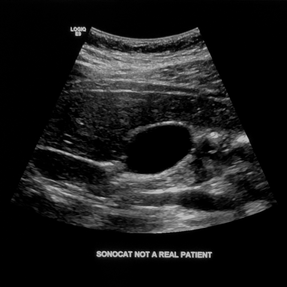
- One image at the body (if normal).
- If the gallbladder is absent, label "Gallbladder Fossa Trans". Cine: Show the neck, body, and fundus.

Gallbladder Trans
Gallbladder Sag/Trans Decub/Erect
- Evaluate in at least one more patient position, repeating the above for gallbladder.
Right Kidney Sag Med/Mid/Lat
- You must show both poles of the kidney clearly. If this is not possible, try different windows or reposition the patient. Color image: Take one image at midpole, showing perfusion in each renal segment.
Measurement image: From upper pole to lower pole not obstructed by bowel.
Cine: Medial to lateral, or lateral to medial.


Rt Kidney Sag
Right Kidney Trans Sup/Mid/Inf
- Transverse kidney should be presented as round and circular as possible, without being too oblique. Color image: Take one image at midpole, showing renal artery and vein at the hilum.
Cine: Superior to inferior.


Rt Kidney Trans
Left Kidney Sag Med/Mid/Lat
- You must show both poles of the kidney clearly. If this is not possible, try different windows or reposition the patient. Color image: Take one image at midpole, showing perfusion in each renal segment.
Measurement image: From upper pole to lower pole not obstructed by bowel.
Cine: Medial to lateral, or lateral to medial.
Left Kidney Trans Sup/Mid/Inf
- Transverse kidney should be presented as round and circular as possible, without being too oblique. Color image: Take one image at midpole, showing renal artery and vein at the hilum.
Cine: Superior to inferior.
Spleen Sag
- Take one image, scanning between the ribs. Measurement image: From diaphragm to inferior edge of the spleen.

Spleen Sag
Spleen Trans
- Take one image, scanning between the ribs. Color image: Take one image at midpole, showing splenic artery and vein at the hilum.

Spleen Trans
Worksheet
- Print the worksheet containing measurements.