Aorta Protocol
- Abdominal aortic aneurysm (AAA) ≥3.0 cm diameter. Scanning Duration: 10 minutes.
Aorta Prox Sag
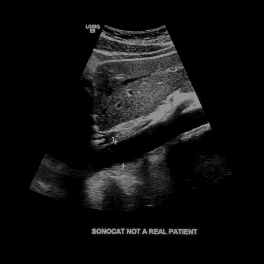
- Show the proximal aorta with the diaphragm in the supine patient position, and take your measurement superior to the celiac trunk. If this view is obstructed by bowel, then scan laterally through the ribs (an example of right lateral approach proximal aorta is shown below).
- Note that in the lateral approach, your sagittal aorta measurement is technically transverse instead of A/P. Therefore, measure the A/P and transverse dimensions of one aorta segment using the same window only. Color image: One optimized image.
Measurement image: Calipers placed from outer wall to outer wall.


Aorta Prox Sag (Anterior and Right Lateral approach)
Aorta Prox Trans
- Note that in the lateral approach, your transverse aorta measurement is technically A/P instead of transverse. Therefore, measure the A/P and transverse dimensions of one aorta segment using the same window only. Color image: One optimized image.
Measurement image: Calipers placed from outer wall to outer wall.

Aorta Prox Trans (Anterior approach)
Aorta Mid Sag
- Show the mid aorta at the level of the renal arteries, just inferior to the SMA. Color image: One optimized image.
Measurement image: Calipers placed from outer wall to outer wall.

Aorta Mid Sag
Aorta Mid Trans
-
Color image: One optimized image.
Measurement image: Calipers placed from outer wall to outer wall.

Aorta Mid Trans
Aorta Distal Sag
- Show the distal aorta before the bifurcation, scanning at the level of the umbilicus. Color image: One optimized image.
Doppler image: Sample the center of the vessel with optimized adjustments.
Measurement image: Calipers placed from outer wall to outer wall.


Aorta Distal Sag
Aorta Distal Trans
-
Color image: One optimized image.
Measurement image: Calipers placed from outer wall to outer wall.

Aorta Distal Trans
Aorta Bifurcation Trans
- In transverse, scan more inferiorly from the distal aorta to show the right and left common iliac artery.
- Your window should be to the left of the umbilicus (patient's left), but use multiple windows and positions as necessary. Color image: One optimized image.

Aorta Bifurcation Trans
Right CIA Sag/Trans
-
Color image: One image each for sagittal and transverse.
Measurement image: Calipers placed from outer wall to outer wall.


Right CIA Sag and Trans
Left CIA Sag/Trans
-
Color image: One image each for sagittal and transverse.
Measurement image: Calipers placed from outer wall to outer wall.


Left CIA Sag and Trans
IVC Prox Sag
- Show the proximal IVC going into the heart.
- Use an anterior or right lateral approach. Color image: One optimized image.
Doppler image: One optimized image.

IVC Prox Sag
Worksheet
- Print the worksheet containing measurements.