Carotid Protocol
- All sagittal images to include gray scale, color, and Doppler.
- When carotid plaque takes up more than 50% of the vessel lumen, sample the velocities directly before and after the plaque, and at the point of most narrowing.
- Ideally, use only one angle of insonation (between 45 and 60 degrees) by steering the Doppler beam rather than changing the angle.
- Always sample the center of the vessel with the Doppler angle parallel to the vessel walls. Scanning Duration: 30 minutes.
Right CCA Prox/Mid/Dist Trans
- (Only the right carotid is demonstrated on this page as an example.)
- Take an image each at the proximal, mid (at the level of thyroid), and distal segments.


Right CCA Trans
Right Bulb/Bifurc Trans
- Take a gray scale and color image each at the carotid bulb and bifurcation. Color image: One image each at the carotid bulb and bifurcation.


Right Bulb and Bifurc Trans
Right CCA Prox/Mid/Dist Sag
- Heel-toe the probe to show the vessel at an angle.
- Show wall-to-wall color filling when there is no plaque, with the color box steered in the correct direction.
- Sample the center of the vessel with adjustments to angle, baseline, speed, scale, and gain. Color image: One image each at the proximal, mid, and distal segments.
Doppler image: Sample the proximal, mid, and distal segments.


Right CCA Sag
Right Bulb Sag
- The probe is angled cranially for the bulb and bifurcation (ICA and ECA).
- The bulb will normally have turbulent flow, so you will see a mix of blue and red color filling. Color image: One optimized image.
Doppler image: One optimized image.


Right Bulb Sag
Right ICA Prox/Mid/Dist Sag
- Follow the sagittal carotid bulb superiorly until you see either the ICA or ECA. Confirm it is the ICA by sampling the Doppler waveform.

- To find a good window and show the elongated ICA, incrementally "crawl" the probe laterally on the neck and angle medially towards the carotid.
- When there is shadowing from calcified plaque, do not take a color image with any gaps in the color filling. If there is no better window, you can over-gain the color to compensate for this, or experiment using B-mode.
- Normal velocity <125 cm/sec. Color image: One image each at the proximal, mid, and distal segments.
Doppler image: Sample the proximal, mid, and distal segments with correct steering.


Right ICA Sag
Right ECA Prox Sag
- Unlike the ICA, the ECA will have branching vessels and a high-resistance waveform.
- When EDV is too small to measure, decrease the wall filter to display very low velocity signals. Color image: One optimized image.
Doppler image: One optimized image.


Right ECA Sag
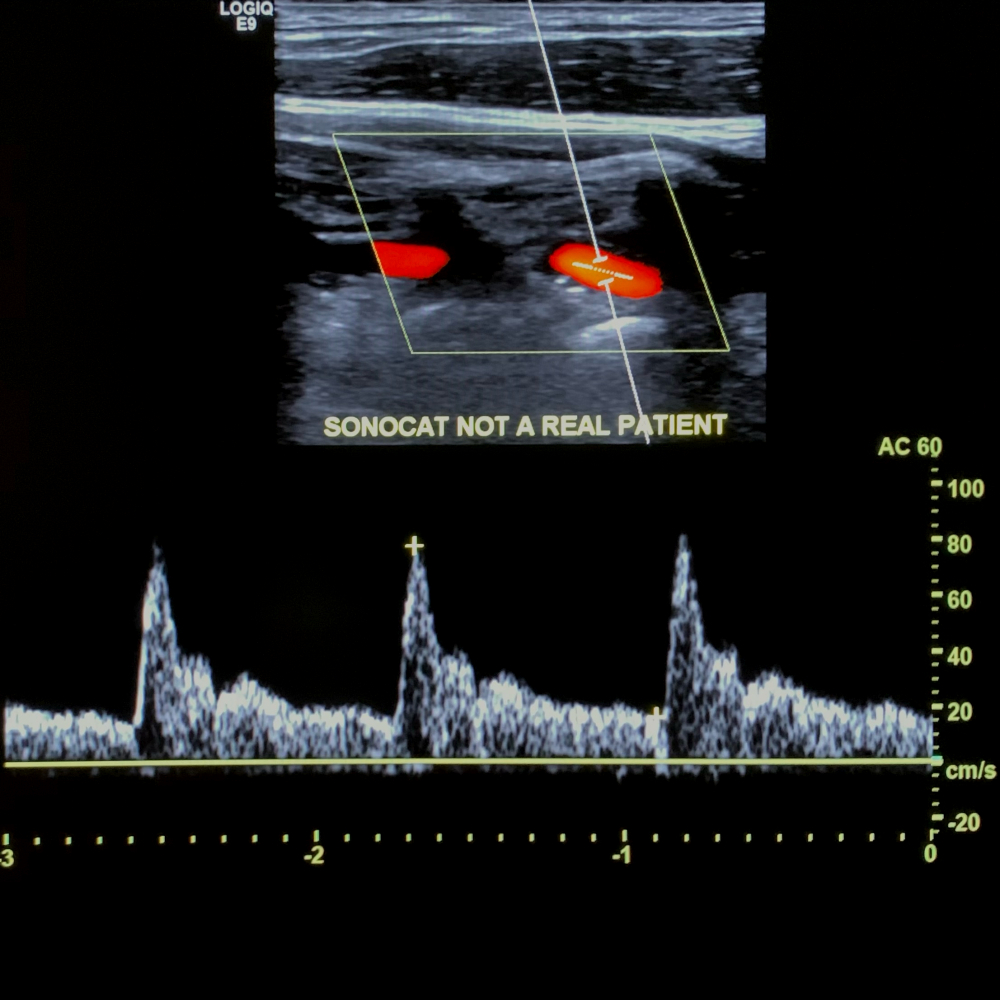
Right Vert Sag
- Start at the sagittal CCA (with or without color), and scan laterally until you see the vertebral artery with the cervical bones.
- Due to the deep position of the vertebral artery, apply more pressure and increase the color gain until there is adequate red color filling.
- You may also see the normal vertebral vein with blue color filling - not to be confused with flow reversal of the vertebral artery, as seen in subclavian steal. Color image: One optimized image.
Doppler image: One optimized image.


Right Vert Sag
Right Vert Sag
- Show the vertebral artery with antegrade flow when it is normal (same direction or color filling as the CCA). Dual-screen image: One image showing both the vertebral artery and ipsilateral CCA with red color filling.

Right Vert Sag
(Repeat the protocol for the left side)
Worksheet
- Print the worksheet containing measurements.