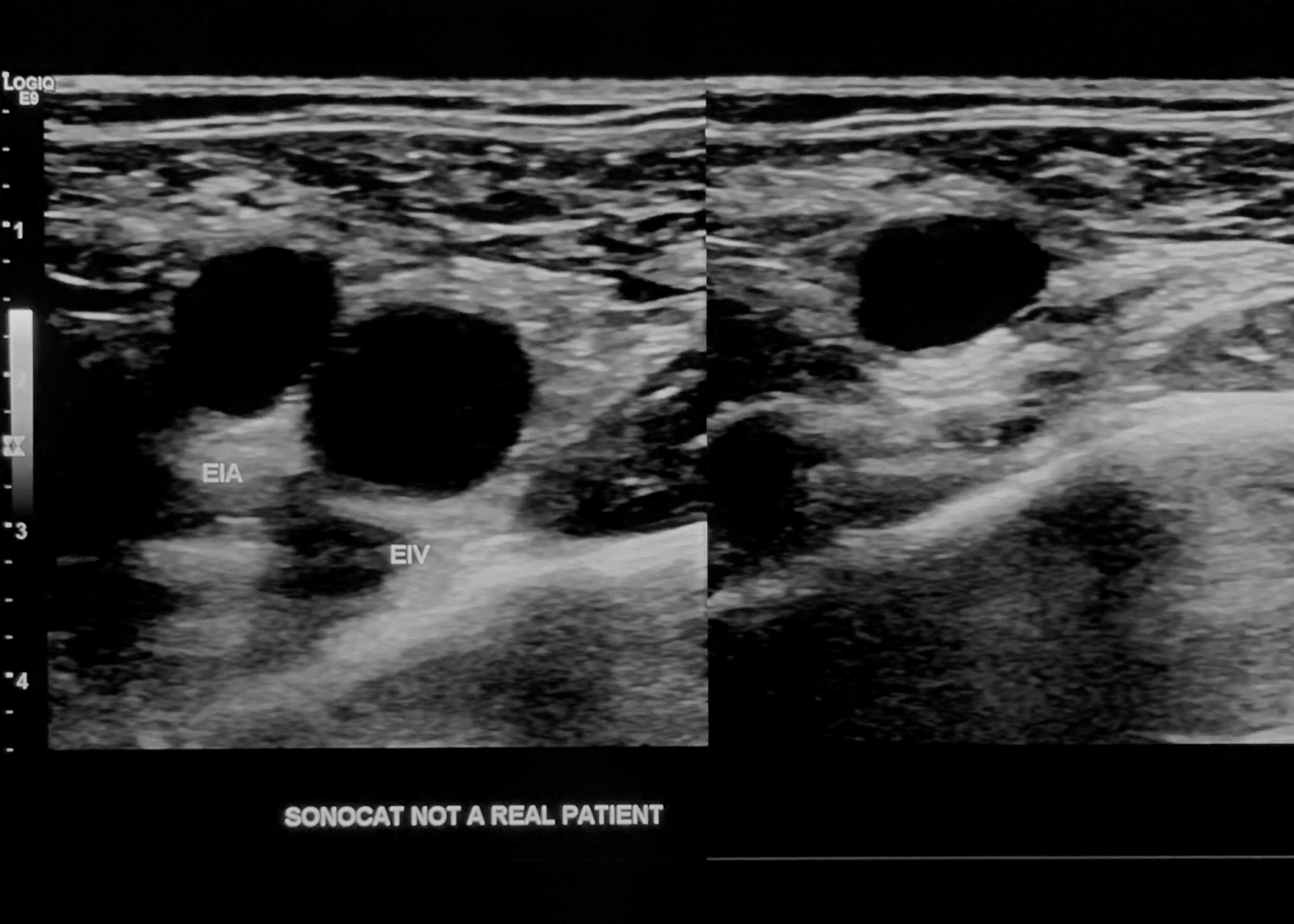
Lower Extremity Venous Protocol
- Gray scale sagittal still images not needed unless positive for thrombus.
- Vein compressions are taken with dual-screen stills above the knee, and cine clips for calf veins. Scanning Duration: 15 minutes for unilateral leg through calf.
Right CFV Trans with compression
- (Only the right leg is demonstrated on this page as an example.)
- Angle your probe cranially and go more superiorly than you would expect.
- You are in the correct position if, by sliding the probe inferiorly, the saphenofemoral junction comes into view. Dual-screen image: One image showing clear compression of CFV.

Right CFV Trans w/ Compression
Right SFJ Trans with compression
- Junction of CFV and greater saphenous vein. Dual-screen image: One image showing clear compression of CFV with GSV.

Right SFJ Trans w/ Compression
Right PFV Prox Trans with compression
- Continue to scan more inferiorly from CFV, and you should see separation of femoral veins and arteries.
- The correct position will have at least four vessels in view (femoral artery, femoral vein, profunda femoris artery, and profunda femoris vein). Dual-screen image: One image showing clear compression of FV prox and PFV.

Right PFV Trans w/ Compression
Right FV Prox Trans with compression
- If your patient is skinny, you may still see the deep femoral vein (aka PFV) as you go inferiorly towards FV prox-mid. Although FV prox is your focus, make sure that the normal PFV also compresses if you catch both veins in your imaging. Dual-screen image: One image showing clear compression of FV prox (and PFV if still seen).

Right FV Prox Trans w/ Compression
Right FV Mid/Dist Trans with compression
- Show distal femoral vein next to the femur bone. Dual-screen image: One image each for FV mid and FV distal showing clear compression.

Right FV Dist Trans w/ Compression
(note the femur bone)
Right CFV Sag
- A normal waveform is phasic with respiration. If there is continuous flow, you should suspect a proximal obstruction e.g. IVC or iliac vein thrombosis, abdominal/pelvic mass, or late stages of pregnancy. Color image: One optimized image.
Doppler image: One optimized image.

Right CFV Sag
Right SFJ Sag
- Sometimes GSV may not be seen if patient is status post varicose vein ablation. Color image: One optimized image.

Right SFJ
Right PFV Prox Sag
- Heel-toe the probe such that the vessels are at an angle as shown below. Color image: One optimized image.

Right PFV Prox Sag
Right FV Prox/Mid/Dist Sag
- Heel-toe the probe such that the vessels are at an angle. Color image: One image each at the proximal, mid, and distal segments.

Right FV Mid Sag
Right Pop V Trans with compression
- Reposition the patient by having him bend his knee and turning the knee outwards. Alternatively, have the patient lie on his side and bend the knee.
- The patient's leg must be relaxed in order to make compression more easy.
- Angle the probe cranially to show one vein (unless duplicated) and one artery.
- Document an incidental popliteal cyst by measuring it, turning on color, and taking cine clips. Dual-screen image: One image showing clear compression of popliteal vein.

Right Pop V Trans w/ Compression
Right Pop V Dist Trans with compression
- Angle your probe inferiorly from previous position until more vessels come into view. Dual-screen image: One image showing clear compression of popliteal vein.

Right Pop V Dist Trans w/ Compression
Right Pop V Sag
- Now have the patient straighten his leg slightly.
- Heel-toe the probe such that the vessels are at an angle. Color image: One optimized image.
Doppler image: One optimized image.

Right Pop V Sag
Right Pop V Dist Sag
- Show the popliteal vein at the trifurcation area. Color image: One optimized image.

Right Pop V Dist Sag
Unilateral vs. Bilateral
- If this is a unilateral leg study, then proceed to evaluate calf veins.
- If this is a bilateral leg study, and your patient has a cancer history, clotting disorder e.g. Factor V Leiden, pulmonary embolus, recent orthopedic surgery or prolonged surgery, pain in this calf, or prior DVT in this calf that needs re-evaluation, then proceed to evaluate calf veins.
- Else (for a bilateral leg study) stop at the knee and repeat the protocol for the contralateral leg.
Right Gastroc V Trans with compression
- Scan the posterior calf to show the paired gastrocnemius veins. Evaluate from superior to inferior, from the popliteal fossa through the gastrocnemius muscle. Dual-screen image: One image showing clear compression of gastrocnemius veins.
Cine: One compression clip.

Right Gastroc V w/ Compression
Right Gastroc V Sag
- Show the paired gastrocnemius veins with color (together in one image or separately).
- Use the slow flow color preset or decrease your color scale.
- Lightly jolt your probe into the patient's calf to promote color filling in the vein. Color image: Images with good color filling.

Right Gastroc V Sag
Right PTV/Peron V Prox/Mid/Dist Trans with compression
- It is imperative to have the patient bend his knee to visualize the peroneal veins properly.
- The correct depth will have the tibia and fibula bones in view. PTVs are found next to the tibia, and peroneal veins are found next to the fibula.
- If needed, switch to a curvilinear probe for deeper penetration. Cine: One compression clip each at the proximal, mid, and distal segments.


Right PTV/Peron V Mid Trans w/ Compression
Right PTV/Peron V Mid Sag
- Show the paired veins with color (together in one image or separately) at the mid calf only when they are normal.
- Use the slow flow color preset or decrease your color scale.
- Lightly jolt your probe into the patient's calf to promote color filling in the vein. Color image: Images with good color filling.

Right PTV/Peron V Mid Sag
Left CFV Sag
- Color and Doppler of the contralateral CFV for unilateral leg exam. Color image: One optimized image.
Doppler image: One optimized image.