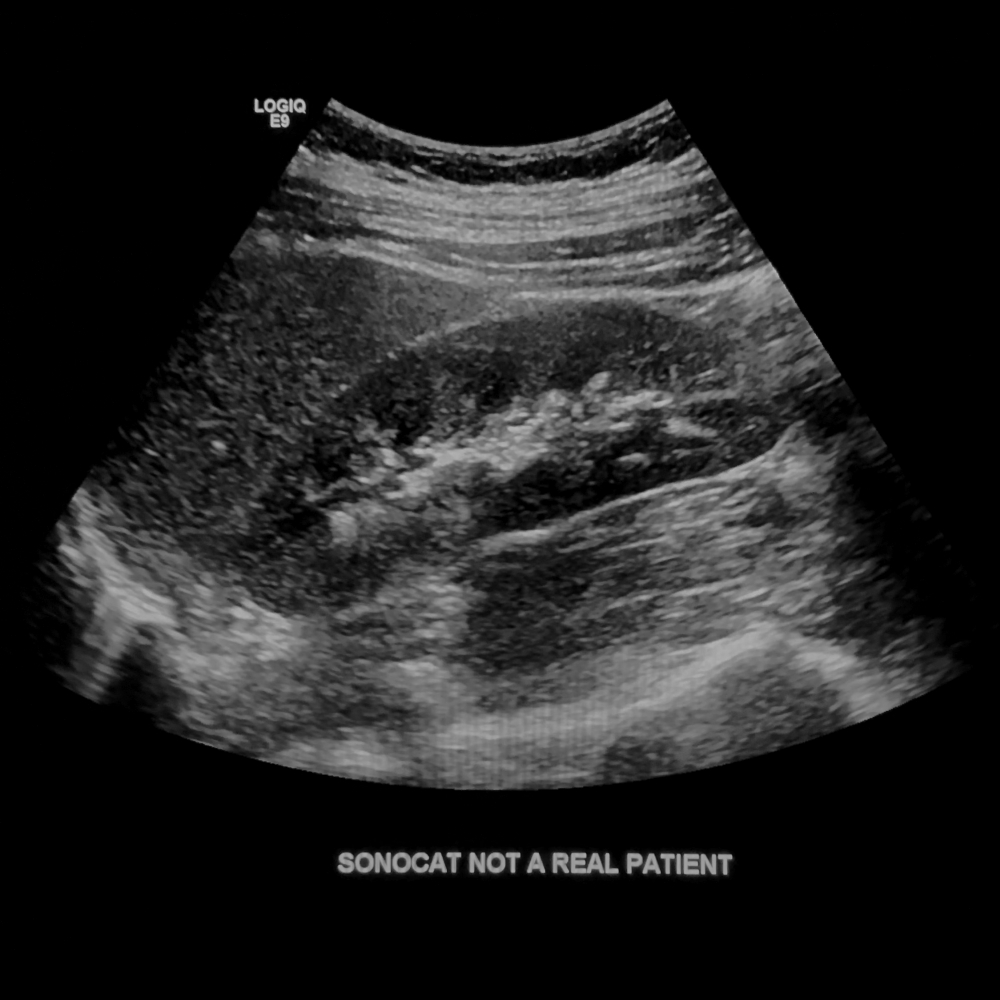
Renal Protocol
- For a septated renal cyst, determine whether the septation has vascularity, and use spectral Doppler if present.
- For hydronephrosis, show the ureteral jet and the post-void kidney for comparison. Scanning Duration: 15 minutes.
Right Kidney Sag
- Take an image showing liver/kidney interface, and measurement.
- To find an optimal window, start in the anterior approach (as shown) with the patient supine, then "crawl" the probe laterally and angle medially towards the kidney. Repeat this motion as needed until you are scanning from the patient's back with the patient lying on his side. Measurement image: From upper pole to lower pole not obstructed by bowel.

Rt Kidney Sag
Right Kidney Sag Med/Mid/Lat
- Show both poles of the kidney clearly. Color image: Take one image at midpole, showing perfusion in each renal segment.
Cine: Medial to lateral, or lateral to medial.


Rt Kidney Sag
Right Kidney Trans Sup/Mid/Inf
- Transverse kidney should be presented as round and circular as possible, without being too oblique. Color image: Take one image at midpole, showing renal artery and vein at the hilum.
Cine: Superior to inferior.


Rt Kidney Trans
Left Kidney Sag
- Take an image showing spleen/kidney interface, and measurement.
- To find an optimal window, start in the anterior approach (as shown) with the patient supine, then "crawl" the probe laterally and angle medially towards the kidney. Repeat this motion as needed until you are scanning from the patient's back with the patient lying on his side. Measurement image: From upper pole to lower pole not obstructed by bowel.

Lt Kidney Sag
Left Kidney Sag Med/Mid/Lat
- Show both poles of the kidney clearly. Color image: Take one image at midpole, showing perfusion in each renal segment.
Cine: Medial to lateral, or lateral to medial.


Lt Kidney Sag
Left Kidney Trans Sup/Mid/Inf
- Transverse kidney should be presented as round and circular as possible, without being too oblique. Color image: Take one image at midpole, showing renal artery and vein at the hilum.
Cine: Superior to inferior.


Lt Kidney Trans
Bladder Trans
- Make note of any intraluminal air within the bladder (a normal finding after cystoscopy), or any layering bladder debris/stone. Reposition your patient to demonstrate the mobility of air or debris/stone.
- In the absence of findings, use TCG to darken the normal bladder lumen or remove artifact, and take one image of the bladder when it is normal.
- You must show the patent ureteral jet when there is hydronephrosis. Cine: Superior to inferior.
Color image: Only when there are findings, or to demonstrate ureteral patency.

Bladder Trans
Bladder Sag
- Take one image of the bladder when it is normal. Cine: Right to left.
Color image: Only when there are findings.

Bladder Sag
Worksheet
- Print the worksheet containing measurements.